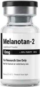
Melanogenesis is a biological process which creates melanin, a category of pigments that provides various colours of skin, hair and eyes. The process of melanogenesis occurs mainly in specialised melanocyte cells, which are located in the basal layer of the epidermis. Even if the process seems easy, it is really complex and consists of several signals, enzymes and interactions.
Melanogenesis is interesting for researchers who work with peptides, as these can interact with biological pathways in a very precise way. Different peptide sequences have already been studied to see their impacts on melanocyte activity, melanin production and the activities of the pathways that play a key role in pigmentation. But it does not mean that all peptides have the same effects. Results can vary considerably depending on the sequence, concentration, experimental model and conditions used.
What happens during melanogenesis?
While the melanocyte cells receive signals that will stimulate melanin synthesis, the process of melanin production starts. One of the main enzymes involved in this process is tyrosinase, an enzyme that catalyses the conversion of the amino acid tyrosine to melanin derivatives.
Concerning melanin, two major types of melanin are usually mentioned, namely eumelanin and pheomelanin. Eumelanin is responsible for black and brown colours, while pheomelanin is responsible for the production of yellowish and reddish colours. The ratio of both pigments during the pigmentation process is mediated by various biological factors, including the transcription factor MITF (microphthalmia-associated transcription factor).
The melanin is produced in special structures referred to as melanosomes. Once formed, these organelles can transfer their pigment content from melanocytes to nearby keratinocytes. The pigment is then distributed through the surrounding epidermal cells.
That makes melanogenesis more than simply a matter of whether tyrosinase is switched on or off. Researchers often look at several points in the pathway when assessing how pigmentation is regulated.
Where do peptides fit into melanogenesis research?
Peptides are of major interest in pigmentary studies due to the influence of their biological action on the amino acid sequence and structure. Particular peptide molecules have been investigated for their functions as signalling compounds, receptor ligands, or as tools for studying melanocyte activity.
An important branch of research focuses on melanocortin signalling. Melanocortins are peptide hormones responsible for many physiological processes, including pigmentation. A crucial peptide in the field of melanogenesis is α-MSH, which is the abbreviation of the term alpha-melanocyte-stimulating hormone.
The interaction of α-MSH with the melanocortin type 1 receptor (MC1R) on the surface of melanocytes has been shown. The activation of MC1R leads to intracellular signalling, which involves an increase in cyclic AMP that directly affects the activity of transcription factors, such as MITF (microphthalmia-associated transcription factor), and enzymes required for melanin production.
Researchers may therefore use peptide-based compounds to investigate how these signalling pathways work, rather than only looking at the final amount of pigment produced.
Tyrosinase and related pathways
Tyrosinase is often used as a key marker in experimental melanogenesis studies because of its central role in melanin synthesis. Researchers may examine tyrosinase expression, enzyme activity or changes in downstream pigmentation markers.
Other proteins are involved as well. Tyrosinase-related protein 1 (TYRP1) and tyrosinase-related protein 2 (TYRP2), also known as dopachrome tautomerase, are part of the broader melanogenic system.
Transcription factors such as MITF (microphthalmia-associated transcription factor) are also important. MITF acts as a regulatory point for several genes involved in melanocyte function and pigment production.
This creates several possible research questions. A study might investigate whether a peptide affects receptor signalling, changes MITF expression, alters tyrosinase activity, or produces a measurable change in melanin content.
Why do experimental models matter?
Melanogenesis research can be carried out using different models, and the choice of model affects the conclusions that can reasonably be drawn.
A biochemical assay yields useful details relevant to understanding the actions of tyrosinase or other enzymes, but while it helps to acquire information about certain processes, it falls short of capturing the full biological nature of melanocytes.
Cellular experiments create a broader context for research. Scientists use the cultured melanocytes and other cell systems to carry out investigations to determine melanin production rates, gene activity and intercell communication.
More sophisticated studies use reconstructed skin or other three-dimensional systems. These models add value but cannot recreate all the aspects of living human skin.
This difference is essential for interpreting scientific findings. A peptide that behaves in an enzyme assay does not prove that the same peptide behaves similarly in a cellular system.
Peptide sequence, purity and study conditions
When investigating peptides, the identity and quality of the material can have a significant bearing on experimental results. Small changes in sequence, modifications or formulation may alter biological behaviour.
Purity is another consideration. A research sample containing impurities, degradation products or incorrect peptide material can complicate interpretation. This is why the scientists use different experimental methods to identify peptides and determine their purity.
It is also important to note that experimental conditions play a significant role. The factors such as concentration of the peptide, time, medium, temperature, and model can affect the results very much.
Specifying this should draw attention to the fact that it mainly refers to a comparison of obtained results between different studies. Two experiments may appear to investigate the same peptide but use different concentrations or biological systems. Their results may therefore not be directly comparable.
What researchers measure?
There is no single measurement that tells the whole story of melanogenesis. Depending on the research question, studies may measure total melanin content, tyrosinase activity, expression of melanogenic genes or levels of signalling molecules.
Microscopy can also be used to observe changes in cellular morphology or pigment distribution. The methods of molecular biology can be useful for the detection of changes in the expression of genes and proteins.
By making various measurements, the specification of logged information makes it possible for the researchers to have a comprehensive picture of the studied phenomenon. For example, an increase in melanin content, together with changes in tyrosinase and MITF, could yield more useful mechanistic information than a pigment measurement alone.
Current research questions
Melanogenesis research continues to examine how pigmentation is regulated at molecular and cellular levels. Peptides are one part of this wider field, alongside small molecules, proteins, hormones and other signalling compounds.
Some areas of interest include receptor-mediated signalling, regulation of melanogenic enzymes, melanosome formation and transfer, oxidative stress and communication between melanocytes and neighbouring skin cells.
There is also growing interest in understanding why pigmentation responses differ between individuals and between different biological conditions. These questions require carefully controlled experiments rather than relying on a single marker or isolated result.
For peptide researchers, therefore, melanogenesis offers a useful model for studying cell signalling and biological activity. The value of the research often lies not simply in whether pigment production increases or decreases, but in understanding how and why a particular peptide changes the pathway.
Reading melanogenesis studies critically
When reviewing research, it is important to verify the experimental model, the identity and concentration of the peptide, the controls, and the methods used to measure pigmentation. A result from an isolated enzyme experiment should be interpreted differently from a result obtained using melanocytes or a reconstructed skin model.
It is also useful to distinguish between evidence showing a biological mechanism and evidence showing an observable outcome. These are related, but they are not interchangeable.
Melanogenesis is a multi-step process, and peptide research within this area reflects that complexity. Looking at the complete signalling pathway can provide a more meaningful understanding than focusing on a single enzyme or a single measurement in isolation.